Abstract
Background
We have shown that the cardiotonic steroid marinobufagenin (MBG) is elevated in clinical and experimental renal disease, and significantly contributes to the development of experimental uremic cardiomyopathy induced by removal of five-sixths of the kidney (5/6 nephrectomy; PNx) in the rat. We have demonstrated that both active and passive immunization against MBG with an anti-MBG monoclonal antibody (mAb 3E9) significantly attenuated cardiac fibrosis following PNx. In the present study we sought to determine whether the use of mAb 3E9 could improve renal function following PNx.
Methods
Sprague–Dawley rats were treated with either mAb 3E9 or with DigiFab (an affinity-purified anti-digoxin antibody formerly named Digibind) during the fourth week after PNx. Sham-operated animals and PNx animals treated with an IgG antibody served as controls. Plasma, urine, and renal tissue were collected at the completion of the study to determine the effects of antibody treatment on renal function.
Results
In PNx rats, treatments with mAb 3E9 and DigiFab, respectively, significantly reduced plasma creatinine, improved creatinine clearance, and reduced proteinuria below the values of these three measures in IgG-treated PNx controls. Additionally, treatment with mAb 3E9 and DigiFab significantly reduced renal fibrosis as measured with Western blotting and Sirius red/Fast green staining.
Conclusions
Passive immunization against MBG significantly improved renal function and markedly reduced renal fibrosis following the experimental induction of renal disease. The work in the study reported here adds to a growing body of knowledge implicating MBG in the development of chronic renal disease. Passive immunization against cardiotonic steroids may serve as a promising treatment for chronic renal disease.
Keywords: blood pressure; cardiotonic steroids; hypertension; marinobufagenin; renal disease; renal fibrosis.
1. Introduction
Elevated levels of endogenous cardiotonic steroids (CTS) have been reported in a variety of clinical conditions associated with plasma volume expansion, such as congestive heart failure (CHF), chronic renal failure, hypertension (HTN), renal ischemia, and preeclampsia.1 We have shown that the cardiotonic steroid marinobufagenin (MBG), signaling through the Na/K-ATPase pathway, causes many of the adverse pathologic effects of experimental renal disease induced by removal of five-sixths of the kidney (5/6 nephrectomy; PNx) in the rat.2,3 Furthermore, our group has demonstrated that pharmacologic administration of MBG causes cardiac hypertrophy and fibrosis in rats, whereas active immunization against MBG attenuated this in PNx animals.2,3 Additionally, treatment of cardiac fibroblasts with MBG at concentrations similar to those reported in experimental and clinical renal failure has been shown to stimulate collagen production.3 This increase in collagen production appears to depend on the Na/K-ATPase-Src-EGFR-ROS signaling cascade.1,3 We have also demonstrated that MBG induces renal fibrosis by stimulating the transcription factor Snail, leading to epithelial-to-mesenchymal transition (EMT) in the tubular epithelia.4
The transcription factor Friend leukemia integration-1 (Fli-1) has been shown to be a negative regulator of collagen synthesis.5,6 Protein kinase C-δ (PKC-δ) phosphorylates Fli-1 and promotes collagen synthesis.7 Our studies indicate that MBG signaling through the Na/K-ATPase-Src-EGFR-ROS cascade initiated PKC-δ translocation to the nucleus, leading to Fli-1 phosphorylation and collagen production.8 Our laboratory recently reported that treatment with a monoclonal antibody (mAb) directed against MBG (3E9 mAb) in PNx animals drastically decreased their blood pressure, significantly reduced cardiac levels of oxidant stress, increased the expression of Fli-1, and significantly reduced cardiac fibrosis.9
The goals of the current study were to determine whether immunization against MBG through the administration of 3E9 mAb would reduce the development of renal fibrosis, and improve renal function in experimental renal failure.
2. Methods
Animal studies
The animal experiments in our study were conducted in accordance with the National Institutes of Health (NIH) Guide for the Care and Use of Laboratory Animals under protocols approved by the University of Toledo Institutional Animal Care and Use Committee. Male Sprague–Dawley rats weighing 250–300 g were used in our studies, with eight sham-nephrectomized rats used as a control group. In 24 animals, PNx was produced by ligation of two-thirds of the arterial blood supply to the left kidney and surgical removal of the right kidney, as reported previously.9 Briefly, rats were anesthetized by inhalation of a mixture of 100% oxygen and 5% isoflurane. Following anesthetization, an incision was made in each animal’s left flank, after which the left kidney of each PNx animal was gently extirpated through the incision so as not to bruise the organ. Following extirpation, the arteries leading to the upper and lower poles of the kidney were ligated and the kidney was observed for characteristic color changes over two-thirds of its surface. One week after the operation on the left kidney, the right kidney was decapsulated to avoid removal of the adrenal gland. After the renal capsule was removed the renal artery, vein, and ureter were ligated and the renal mass above the ligature was removed. This maneuver produces sustained hypertension (HTN) within 2 weeks and significant cardiac fibrosis and hypertrophy by 4 weeks after surgery.2,3,9
Ar 4 weeks after surgical manipulation, rats were given the following via intraperitoneal injections: IgG (n=8) used as a negative control for 3E9 mAb immunization; DigiFab (n=8), or 3E9 mAb against MBG (n=8), with this mAb provided by the laboratory of Drs. Alexei Bagrov and Olga Fedorova of the National Institute on Aging. DigiFab was administered at a dose of 10 µg/kg, which was similar to the doses of DigiFab used in patients with preeclampsia,10,11 and was the same dose that we have shown to reduce cardiac fibrosis following PNx.9 The dose of 3E9 mAb was 50 µg/, as shown previously by our group to reduce cardiac fibrosis and provide a sustained blood pressure effect in rats subjected to PNx.9 One week antibody treatment (5 weeks following PNx) with 3E9 mAb, the rats were euthanized and the remnant kidneys were collected. Immediately retrieval of each remnant kidney, it was dissected along the apical midline, resulting in a bivalve cut of the kidney. One half of the kidney was immediately frozen in liquid nitrogen and stored at –80 °C for use in biochemical analysis, and the other half was fixed in 4% formalin- buffered solution (pH 7.2) for 18 hours and then dehydrated in 70% ethanol. Dehydration was followed by embedding in paraffin and cutting with a microtome.
Creatinine and creatinine clearance
At the conclusion of the study, 24-hour urine samples were collected. Following the urine collection the study animals were euthanized and blood samples were obtained from the abdominal aorta. Plasma and urine creatinine were evaluated with a colorimetric method using a commercial kit from Teco Diagnostics (Anaheim, CA) according to the manufacturer’s protocol, as we have previously described.9 Creatinine clearance was calculated as previously described.9
Western Blot analyses of collagen-1
Western blot analyses were doneon proteins from tissue homogenates as previously described.3,9 Briefly, kidney tissue was homogenized in ice-cold radioimmunoprecipitaton (RIPA) lysis buffer (pH 7.0; Santa Cruz Biotechnology, Santa Cruz, CA). The homogenate was centrifuged at 1,400 × g for 30 seconds at 4 °C. The supernatant was discarded and the pellet fraction was resuspended in 5% sodium dodecyl sulfate (SDS) in 50 mmol/l Tris-HCl (pH 7.4). The protein in the in the resuspended pellet fraction was quantified and was solubilized at a concentration of 10–20 µg of protein per well in sample buffer (2% SDS, 5% β-mercaptoethanol, 20% glycerol, 0.005% bromophenol blue, and 50 mmol/l Tris-HCl; pH 7.0). The components of the protein were then resolved via SDS–polyacrylamide gel electrophoresis (PAGE), using precast Ready Gels 4%–15% Tris-HCl, purchased from Bio-Rad (Hercules, CA). Following PAGE, the proteins were electrotransferred from the gel onto nitrocellulose membranes. The membranes were blocked with 5% nonfat dry milk in Tris-HCl at 20 mmol/l (pH 7.5, 150 mmol/l NaCl and 0.1% Tween-20). Goat anti-type-1 collagen antibody (Southern Biotech, Birmingham, AL) was used to probe for collagen-1, and a secondary horseradish peroxidase-conjugated anti-goat antibody was purchased from Santa Cruz Biotechnology. Chemiluminescent detection and quantification were done with ECL and ECL-Plus reagents (Amersham Biosciences, Ronkonkoma, MA). Loading conditions were controlled with a polyclonal anti-actin goat antibody (Santa Cruz Biotechnology).
Histology
Sirius red/Fast Green staining for collagen was performed on experimentally treated renal tissues and fibrosis was quantified through a protocol modeled after Junquiera and Puchtler.12,13 Briefly, 4-µm sections of paraffin-embedded renal tissues were mounted on slides and subsequently deparaffinized by overnight incubation in xylene, after which the tissue sections were re-hydrated by sequential 3-minute incubations in 100% ethanol (EtOH, 3x), 95% EtOH (1x), and 80% EtOH (1x). The slides were then incubated for 1 hour in Sirius red solution (0.25g, Direct Red 80, Sigma; 250 ml saturated aqueous picric acid, RICCA Chemical, Arlington, TX; and 250 µl picric acid reagent plus, Sigma). Following this, the slides were washed in three changes of acidified water (0.005% acetic acid solution) and then incubated in Fast Green dying solution (0.25g, Fast Green FCF, Sigma; 250 ml saturated aqueous picric acid; and 250 µl picric acid reagent plus) for 1 hour. The slides were then washed again in three changes of acidified water, dehydrated in 3 changes of 100% EtOH, cleared in xylene, and mounted with a resinous medium. Brightfield images of the stained slides were made at a magnification of 20 x on an FSX100 system (Olympus, Tokyo, Japan) and analyzed for collagen by ImageJ image-processing software (US National Institutes of Health).
Proteinuria
The protein concentrations in the 24-hour urine samples collected at the end of the experiment were determined and urine creatinine was measured as described above. The protein concentrations in the urine samples were measured with the Bio-Rad DC Protein assay according to the manufacturer’s protocol (Bio-Rad). Urinary protein concentrations are normalized for urine creatinine excretion.
Statistical analyses
The results of analyses of the study data are presented as means ± standard error of the mean (SEM). Data were analyzed through one-way analysis of variance followed by the Newman–Keuls test (for intragroup analyses), through repeated measures analysis of variance followed by the Newman–Keuls test (for intergroup analyses), and with two tailed t-tests when applicable (GraphPad Prism Software, San Diego, CA). A two-sided value of P < 0.05 was considered statistically significant.
3. Results
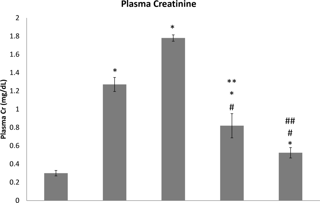
Sprague–Dawley rats subjected to PNx display hypertension, fourfold increases in their MBG concentration, marked increases in plasma creatinine, and systemic oxidative stress, as illustrated previously by our study group.9 In addition to these effects, these animals develop cardiac hypertrophy and fibrosis, as we have also previously shown,2,3,9 and demonstrate significant renal failure, as seen in the present study. Figure 1 illustrates a substantial increase in plasma creatinine in rats subjected to PNx as opposed to sham-operated controls (1.27 ± 0.08 mg/dl vs. 0.30 ± 0.03 mg/dl, P < 0.01).
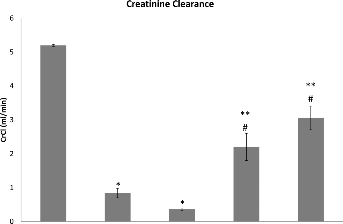
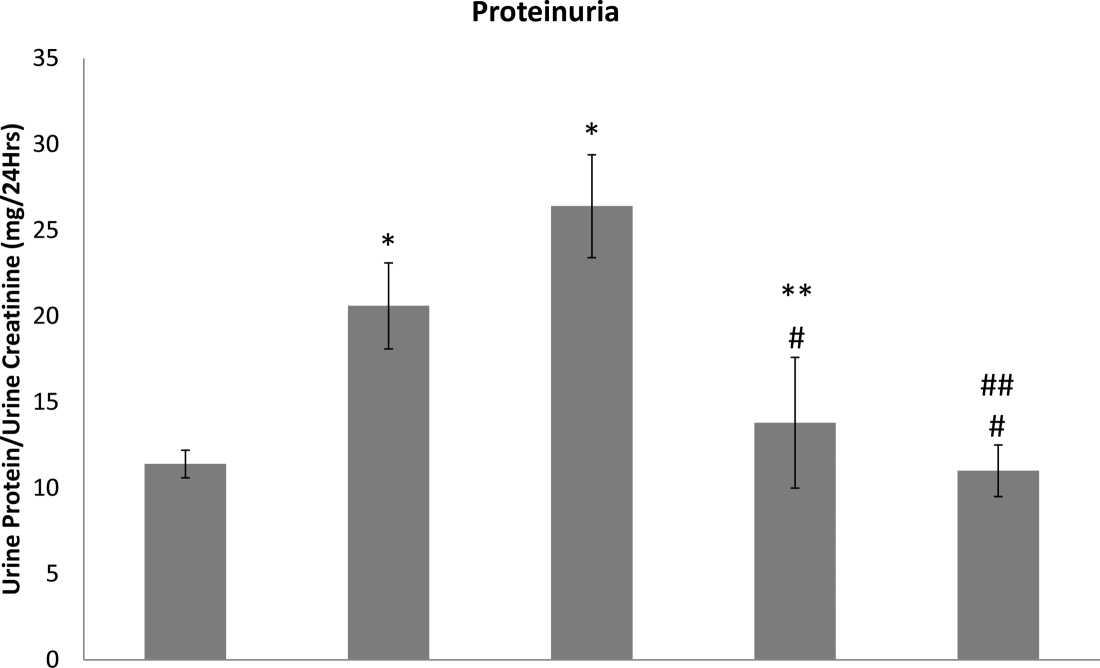
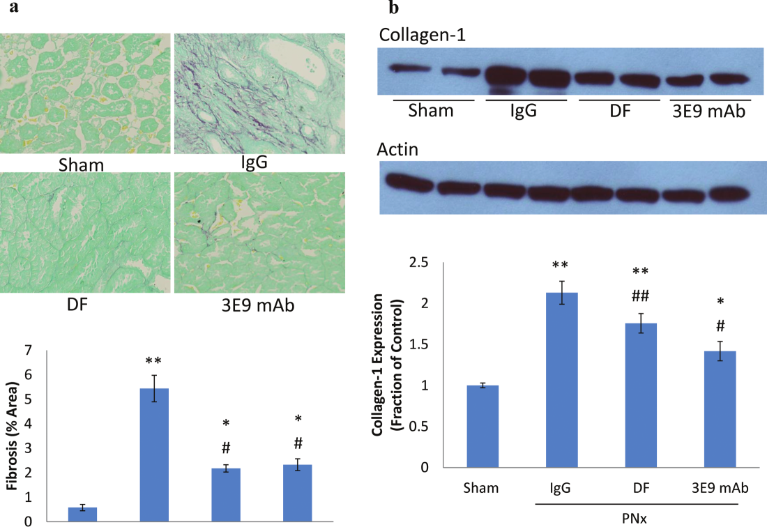
Additionally, PNx animals displayed significantly reduced creatinine clearance (0.84 ± 0.14 ml/min vs. 5.2 ± 0.03 ml/min, P < 0.01) (Figure 2), as well as significantly greater proteinuria than did sham-operated controls (20.6 ± 2.5 mg/24 h vs. 11.4 ± 0.8 mg/24 h, P < 0.01) (Figure 3). Partial nephrectomy induced an almost sixfold increase in kidney fibrosis as compared with sham-operated controls, as measured with Sirius Red staining (P < 0.01, Figure 4a), as well as a twofold increase in collagen-1 protein levels in renal tissue (P < 0.01, Figure 4b) as compared with sham-operated controls. These results reveal the significant nature of the renal failure and injury elicited by PNx.
Immunization with either DigiFab (0.82 ± 0.13 mg/dl vs. 1.78 ± 0.04 mg/dl, P < 0.01) (Figure 1), or 3E9 mAb (0.52 ± 0.06 mg/dl vs. 1.78 ± 0.04 mg/dl, P < 0.01) (Figure 1) significantly reduced plasma creatinine cooncentrations as compared with those of PNx animals given IgG antibody. In addition, treatment with 3E9 mAb (3.06 ± 0.35 ml/min vs. 0.36 ± 0.04 ml/min, P < 0.01) (Figure 2) or DigiFab (2.20 ± 0.40 vs. 0.36 ± 0.04 ml/min, P < 0.01) (Figure 2) at 4 weeks after PNx significantly increased creatinine clearance above that of animals treated with IgG. Although the restoration of creatinine clearance with 3E9 mAb and DigiFab was still significantly smaller than that observed for sham-operated rats, it does represent a 2.5- to 3-fold increase over that observed with PNx or PNx followed by IgG (Figure 2).
As compared with that in IgG-treated animals, proteinuria was also significantly reduced in animals that received 3E9 mAb (11.0 ± 1.5 vs. 26.4 ± 3.0 mg/24h, P < 0.01) (Figure 3) and in those that received DigiFab (13.8 ± 3.8 mg/24 h vs. 26.4 ± 3.0 mg/24h, P < 0.01) (Figure 3). This almost twofold decrease in proteinuria resulted in levels of proteinuria similar to those observed in sham-operated controls (Figure 3).
Lastly, injection of either 3E9 mAb or DigiFab at 4 weeks after PNx reduced renal injury. Kidney fibrosis as measured with Sirius red/Fast Green staining was reduced by twofold in both DigiFab-treated (P < 0.01, Figure 4a) and 3E9 mAb–treated animals (P < 0.01, Figure 4a) as compared with that in IgG-treated animals, although the level of fibrosis in the DigiFab-treated and 3E9 mAb-treated animnals was still higher than that observed in sham-treated rats (Figure 4a). Renal collagen-1 protein expression was significantly reduced in 3E9 mAb- (P < 0.01, Figure 4b) and DigiFab-treated animals (P < 0.05, Figure 4b) as compared with those treated with IgG. This reduction resulted in collagen-1 levels in kidney tissue that were still significantly higher than those in sham operated animals. These results suggest that immunoneutralization of CTS significantly eases the loss of renal function induced by experimental renal failure attributable to increased circulating levels of CTS.
4. Discussion
Our previous investigation of the ability of passive immunization against MBG to ease the deleterious effects of PNx on cardiovascular function and injury revealed that immunization with 3E9 mAb and Digibind reversed cardiac fibrosis and hypertrophy in animals subjected to PNx followed by immunization against CTS.9 The present study has shown that a single administration of an mAb against an endogenous Na/K-ATPase inhibitor, MBG, in rats with experimentally induced renal failure dramatically improved these animals’ kidney function, as illustrated by increased creatinine clearance, decreased plasma creatinine levels, decreased proteinuria, and a significant reduction in renal fibrosis. Indeed, animals subjected to PNx and treated after the onset of renal injury with either 3E9 mAb or DigiFab, a commercially used antibody against CTS, had dramatically improved kidney function as compared with PNx animals treated with IgG.
Additionally, the rats injected with antibodies to CTS also showed significantly less renal injury than either PNx animals or PNx animals treated with IgG. These findings, together with our previous findings that injection of 3E9 mAb resulted in a sustained amelioration of blood pressure, reduced cardiac fibrosis, and reduced collagen synthesis in cardiac tissues of rats subjected to PNx or to PNx followed by the administration of IgG, demonstrate the potential importance of CTS in the progression of kidney and cardiac disease in experimental renal failure in the rat.9
Previous studies have revealed that remnant kidney fibrosis plays a role in the progression of renal failure in the PNx rat model used in the present study.14,15 This study provides renal morphologic and biochemical data illustrating that a single injection of either 3E9 mAb or DigiFab reversed the progressive kidney disease observed in animals subjected to PNx and PNx followed by the administration of IgG. The study also demonstrates that kidney function is dramatically improved with a single administration of anti-CTS antibodies. This study, together with our previous findings that immunoneutralization with MBG eases cardiac fibrosis and has a pressor effect in partial nephrectomy, illustrates the need for continued studies of the role of CTS in the pathogenesis of renal fibrosis.9
Acknowledgments
Steven T. Haller is supported by the National and Ohio Valley Affiliate of the American Heart Association (13POST16860035). Joseph I. Shapiro and Jiang Tian have received support from the National Institutes of Health (HL109015, HL071556, and HL105649).
Disclosure
The authors declare no conflicts of interest.
References
| # | Year | Authors | Title |
|---|---|---|---|
| 1 | 2009 | Bagrov AY, Shapiro JI, Fedorova OV | Endogenous cardiotonic steroids: physiology, pharmacology, and novel therapeutic targets. Pharmacol Rev 2009; 61:9–38. |
| 2 | 2006 | Kennedy DJ, Vetteth S, Periyasamy SM, Kanj M, Fedorova L, Khouri S, Kahaleh MB, Xie Z, Malhotra D, Kolodkin NI, Lakatta EG, Fedorova OV, Bagrov AY, Shapiro JI | Central role for the cardiotonic steroid marinobufagenin in the pathogenesis of experimental uremic cardiomyopathy. Hypertension 2006; 47:488–495. |
| 3 | 2007 | Elkareh J, Kennedy DJ, Yashaswi B, Vetteth S, Shidyak A, Kim EG, Smaili S, Periyasamy SM, Hariri IM, Fedorova L, Liu J, Wu L, Kahaleh MB, Xie Z, Malhotra D, Fedorova OV, Kashkin VA, Bagrov AY, Shapiro JI | Marinobufagenin stimulates fibroblast collagen production and causes fibrosis in experimental uremic cardiomyopathy. Hypertension 2007; 49:215–224. |
| 4 | 2009 | Fedorova LV, Raju V, El-Okdi N, Shidyak A, Kennedy DJ, Vetteth S, Giovannucci DR, Bagrov AY, Fedorova OV, Shapiro JI, Malhotra D | The cardiotonic steroid hormone marinobufagenin induces renal fibrosis: implication of epithelial-to-mesenchymal transition. Am J Physiol Renal Physiol 2009; 296:F922–934. |
| 5 | 2001 | Czuwara-Ladykowska J, Shirasaki F, Jackers P, Watson DK, Trojanowska M | Fli-1 inhibits collagen type i production in dermal fibroblasts via an sp1-dependent pathway. J Biol Chem 2001; 276:20839–20848. |
| 6 | 2006 | Wang Y, Fan PS, Kahaleh B | Association between enhanced type i collagen expression and epigenetic repression of the fli1 gene in scleroderma fibroblasts. Arthritis Rheum 2006; 54:2271–2279. |
| 7 | 2005 | Jinnin M, Ihn H, Yamane K, Mimura Y, Asano Y, Tamaki K | Alpha2(i) collagen gene regulation by protein kinase c signaling in human dermal fibroblasts. Nucleic Acids Res 2005; 33:1337–1351. |
| 8 | 2009 | Elkareh J, Periyasamy SM, Shidyak A, Vetteth S, Schroeder J, Raju V, Hariri IM, El-Okdi N, Gupta S, Fedorova L, Liu J, Fedorova OV, Kahaleh MB, Xie Z, Malhotra D, Watson DK, Bagrov AY, Shapiro JI | Marinobufagenin induces increases in procollagen expression in a process involving protein kinase c and fli-1: implications for uremic cardiomyopathy. Am J Physiol Renal Physiol 2009; 296:F1219–1226. |
| 9 | 2012 | Haller ST, Kennedy DJ, Shidyak A, Budny GV, Malhotra D, Fedorova OV, Shapiro JI, Bagrov AY | Monoclonal antibody against marinobufagenin reverses cardiac fibrosis in rats with chronic renal failure. Am J Hypertens 2012; 25:690–696. |
| 10 | 1988 | Goodlin RC | Antidigoxin antibodies in eclampsia. N Engl J Med 1988; 318:518–519. |
| 11 | 1996 | Adair CD, Buckalew V, Taylor K, Ernest JM, Frye AH, Evans C, Veille JC | Elevated endoxin-like factor complicating a multifetal second trimester pregnancy: treatment with digoxin-binding immunoglobulin. Am J Nephrol 1996; 16:529–531. |
| 12 | 1979 | Junquiera LC, Junqueira LC, Brentani RR | A simple and sensitive method for the quantitative estimation of collagen. Anal Biochem 1979; 94:96–99. |
| 13 | 1973 | Puchtler H, Waldrop FS, Valentine LS | Polarization microscopic studies of connective tissue stained with picro-sirius red fba. Beitr Pathol 1973; 150:174–187. |
| 14 | 2009 | An WS, Kim HJ, Cho KH, Vaziri ND | Omega-3 fatty acid supplementation attenuates oxidative stress, inflammation, and tubulointerstitial fibrosis in the remnant kidney. Am J Physiol Renal Physiol 2009; 297:F895–903. |
| 15 | 2011 | Sun L, Zhang D, Liu F, Xiang X, Ling G, Xiao L, Liu Y, Zhu X, Zhan M, Yang Y, Kondeti VK, Kanwar YS | Low-dose paclitaxel ameliorates fibrosis in the remnant kidney model by down-regulating mir-192. J Pathol 2011; 225:364–377. |